
Genomics of Liver Tumors
Jessica
ZUCMAN-ROSSI
We study 3 types of solid tumors using genomic approaches to better understand their molecular and clinical heterogeneity and develop new diagnostic and prognostic markers. For this we use the last advanced technologies in molecular biology associated to our expertise in bioinformatic.
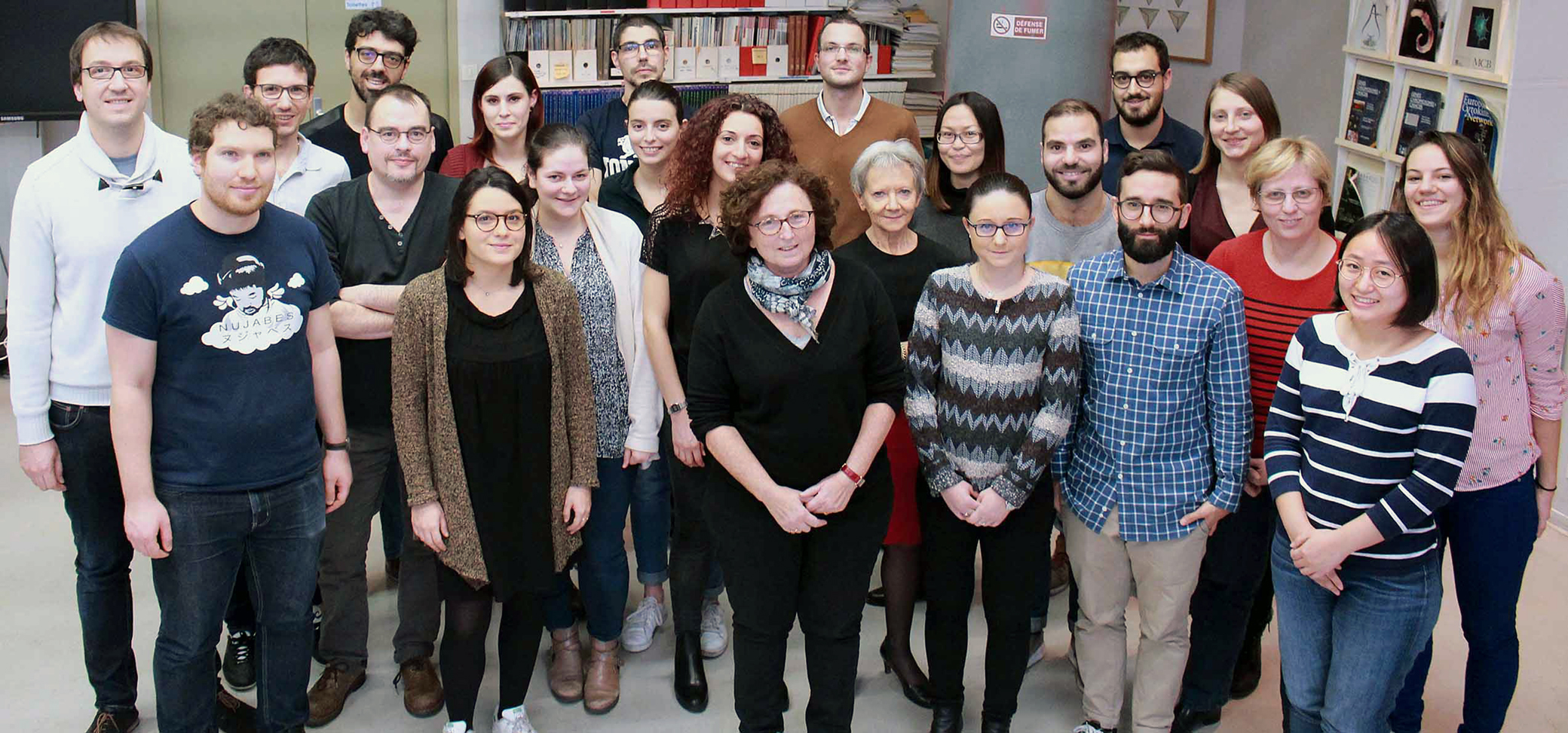
The various activities of our lab and its members
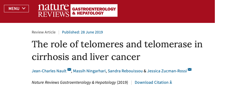
Telomerase is a key enzyme for cell survival that prevents telomere shortening and the subsequent cellular senescence that is observed after many rounds of cell division. In contrast, inactivation of telomerase is observed in most cells of the adult liver. Absence of telomerase activity and shortening of telomeres has been implicated in hepatocyte senescence and the development of cirrhosis, a chronic liver disease that can lead to hepatocellular carcinoma (HCC) development. During hepatocarcinogenesis, telomerase reactivation is required to enable the uncontrolled cell proliferation that leads to malignant transformation and HCC development. Part of the telomerase complex, telomerase reverse transcriptase, is encoded by TERT, and several mechanisms of telomerase reactivation have been described in HCC that include somatic TERT promoter mutations, TERTamplification, TERT translocation and viral insertion into the TERTgene. An understanding of the role of telomeres and telomerase in HCC development is important to develop future targeted therapies and improve survival of this disease. In this Review, the roles of telomeres and telomerase in liver carcinogenesis are discussed, in addition to their potential translation to clinical practice as biomarkers and therapeutic targets.
Eric Letouzé and Stefano Caruso from our lab will present their works tomorrow at the CRC Scientific Day!
Don’t miss this day with high level science from our Research Center !
More informations on CRC website:
The Faculty of Medicine and Dentistry of UC Louvain will present the honoris causa doctor insignia today, May 29 2019 to our Director, Pr. Jessica Zucman-Rossi. A new prestigious reward for our director.

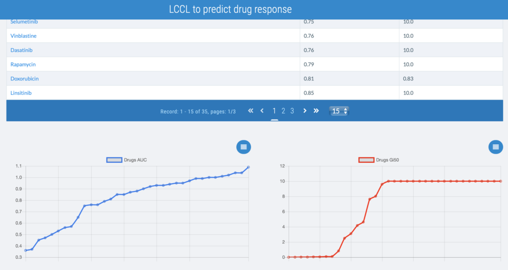
The last work of Sandra Rebouissou group is newly accepted for publication in Gastroenterology.
Entitled, “Analysis of Liver Cancer Cell Lines Identifies Agents With Likely Efficacy Against Hepatocellular Carcinoma and Markers of Response” it is the first study to provide a comprehensive molecular characterization of most widely used liver cancer cell lines. All the data are available at : www.lccl.zucmanlab.com
Article available on Gastroenterology website.
Cyclins A2 and E1 regulate the cell cycle by promoting S phase entry and progression. Here, we identify a hepatocellular carcinoma (HCC) subgroup exhibiting cyclin activation through various mechanisms including hepatitis B virus (HBV) and adeno-associated virus type 2 (AAV2) insertions, enhancer hijacking and recurrent CCNA2 fusions. Cyclin A2 or E1 alterations define a homogenous entity of aggressive HCC, mostly developed in non-cirrhotic patients, characterized by a transcriptional activation of E2F and ATR pathways and a high frequency of RB1 and PTEN inactivation. Cyclin-driven HCC display a unique signature of structural rearrangements with hundreds of tandem duplications and templated insertions frequently activating TERT promoter. These rearrangements, strongly enriched in early-replicated active chromatin regions, are consistent with a break-induced replication mechanism. Pan-cancer analysis reveals a similar signature in BRCA1-mutated breast and ovarian cancers. Together, this analysis reveals a new poor prognosis HCC entity and a rearrangement signature related to replication stress.
We recently identified a novel histological subtype of hepatocellular carcinoma, designated as "macrotrabecular-massive" (MTM-HCC) and associated with specific molecular features. In order to assess the clinical relevance of this novel variant, we aimed to investigate its prognostic value in two large series of patients with HCC treated either by surgical resection or radiofrequency ablation (RFA). We retrospectively included 237 HCC surgical samples and 284 HCC liver biopsies from patients treated by surgical resection and RFA, respectively. Histological slides were reviewed by pathologists specialized in liver disease, and the MTM-HCC subtype was defined by the presence of a predominant (>50%) macrotrabecular architecture (more than 6 cells thick). The main clinical and biological features were recorded at baseline. Clinical endpoints were early and overall recurrence. The MTM-HCC subtype was identified in 12% of the whole cohort (16% of surgically resected samples, 8.5% of liver biopsy samples). It was associated at baseline with known poor prognostic factors (tumor size, AFP level, satellite nodules and vascular invasion). Multivariate analysis showed that MTM-HCC subtype was an independent predictor of early and overall recurrence (surgical series: OR 3.03 (1.38-6.65), p=0.006 and 2.76 (1.63-4.67), p<0.001); RFA series: 2.37 (1.36-4.13), p=0.002 and 2.19 (1.35-3.54), p=0.001, respectively). Its prognostic value was retained even after patients stratification according to common clinical, biological and pathological features of aggressiveness. No other baseline parameter was independently associated to recurrence in the RFA series. The MTM-HCC subtype, reliably observed in 12% of patients eligible for a curative treatment, represents an aggressive form of HCC that may require more specific therapeutic strategies.
Genomic alterations driving tumorigenesis result from the interaction of environmental exposures and endogenous cellular processes. With a diversity of risk factors, liver cancer is an ideal model to study these interactions. Here, we analyze the whole genomes of 44 new and 264 published liver cancers and we identify 10 mutational and 6 structural rearrangement signatures showing distinct relationships with environmental exposures, replication, transcription, and driver genes. The liver cancer-specific signature 16, associated with alcohol, displays a unique feature of transcription-coupled damage and is the main source of CTNNB1 mutations. Flood of insertions/deletions (indels) are identified in very highly expressed hepato-specific genes, likely resulting from replication-transcription collisions. Reconstruction of sub-clonal architecture reveals mutational signature evolution during tumor development exemplified by the vanishing of aflatoxin B1 signature in African migrants. Finally, chromosome duplications occur late and may represent rate-limiting events in tumorigenesis. These findings shed new light on the natural history of liver cancers.
Hepatocellular adenomas (HCAs) are benign liver tumors that can be assigned to molecular subtypes based on inactivating mutations in hepatocyte nuclear factor 1A, activating mutations in β-catenin, or activation of inflammatory signaling pathways. We aimed to update the classification system for HCA and associate the subtypes with disease risk factors and complications. We analyzed expression levels of 20 genes and sequenced exon regions of 8 genes (HNF1A, IL6ST, CTNNB1, FRK, STAT3, GNAS, JAK1, and TERT) in 607 samples of 533 HCAs from 411 patients, collected from 28 centers mainly in France from 2000 and 2014. We performed gene expression profile, RNA sequence, whole-exome and genome sequence, and immunohistochemical analyses of select samples. Molecular data were associated with risk factors, histopathology, bleeding, and malignant transformation. Symptomatic bleeding occurred in 14% of the patients (85% of cases were female, median age, 38 years); 7% of the nodules were borderline between HCA and hepatocellular carcinoma, and 3% of patients developed hepatocellular carcinoma from HCA. Based on molecular features, we classified HCA into 8 subgroups. One new subgroup, composed of previously unclassified HCA, represented 4% of HCAs overall and was associated with obesity and bleeding. These tumors were characterized by activation of sonic hedgehog signaling, due to focal deletions that fuse the promoter of INHBE with GLI1. Analysis of genetic heterogeneity among multiple HCAs, from different patients, revealed a molecular subtype field effect; multiple tumors had different mutations that deregulated similar pathways. Specific molecular subtypes of HCA associated with various HCA risk factors, including imbalances in estrogen or androgen hormones. Specific molecular subgroup of HCA with β-catenin and sonic hedgehog activation associated with malignant transformation and bleeding, respectively....
Hepatocellular carcinomas (HCCs) are liver tumors related to various etiologies, including alcohol intake and infection with hepatitis B (HBV) or C (HCV) virus. Additional risk factors remain to be identified, particularly in patients who develop HCC without cirrhosis. We found clonal integration of adeno-associated virus type 2 (AAV2) in 11 of 193 HCCs. These AAV2 integrations occurred in known cancer driver genes, namely CCNA2 (cyclin A2; four cases), TERT (telomerase reverse transcriptase; one case), CCNE1 (cyclin E1; three cases), TNFSF10 (tumor necrosis factor superfamily member 10; two cases) and KMT2B (lysine-specific methyltransferase 2B; one case), leading to overexpression of the target genes...


